-
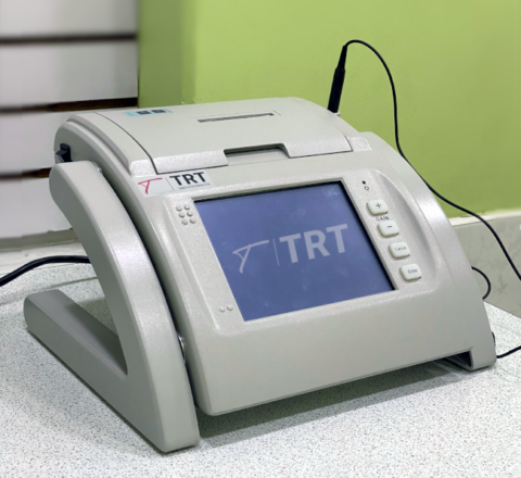

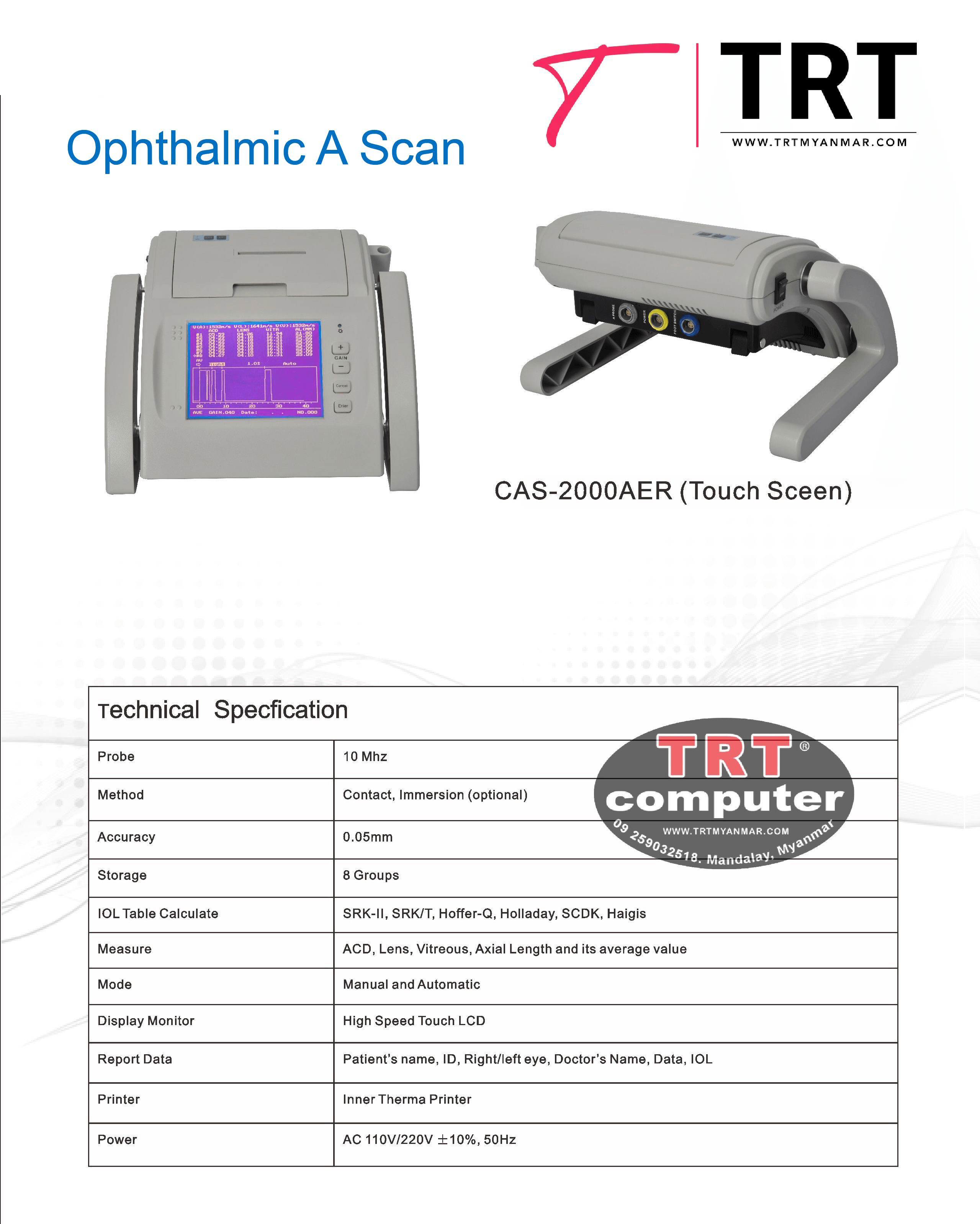
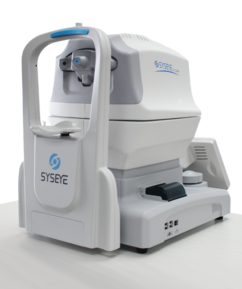
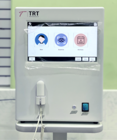
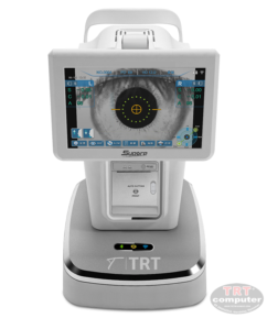
OPHTHALMIC A SCAN Touch Screen
- Inner thermal printer
- High speed LCD, Touch screen
- Report Data: Patient’s name, ID, Right/Left, Doctor’s name, Date, IOL Table
- Modes: Manual , Automatic
-

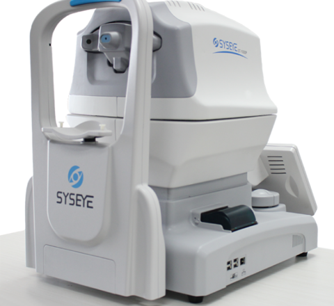
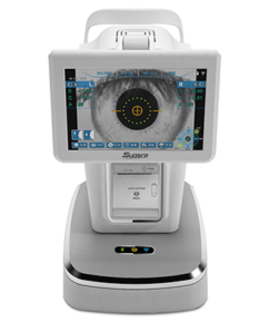
Product features:
Auto tonometer utilizes image control and feature recognition for auto 3-D positioning of XYZ with reliable judgment and measurement results, and easy to operate. The air non-contact approach to measure IOP can also be used for corneal thickness (ST-1000P).
ST-1000 Tonometer only
ST-1000P tonometer with Pachymeter• Auto 3-D positioning of XYZ results in precise and reliable measurement
• Full-auto measurement for both right and left eye data without manual switch
• Easy to operate the tonometer with color touch monitor
• Stable data of 10P through soft air measurement
• To compensate corneal thickness measurement of IOP -

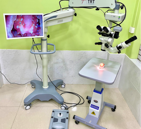
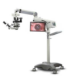
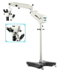
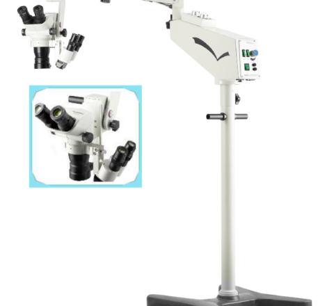
Features:
- Generous and beautiful design, convenient installation and humanized operation;
- Superior quality Japanese OLYMPUS SZX7 Main Microscope;
- Adaptable with video system to record operations for teaching and study purposes;
- Independent stereo coaxial Assistant Microscope;
- DO The surgery is safer by adopted aspherical illumination system and red reflex set ;
- Stable and reliable X-Y speed, with imported low noise motor; Accurate location and fine control performance;
- Natural color, clear stereo field of view, and high resolution contrast images;
- The operation is convenient by double bulbs design and compact panel;
- OO There are 8 functions in one Foot Switch to meet different surgery needs;
- BIOM Lens & Image Inverting Lens can be used to meet retinal vitreous surgery;
-
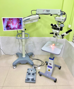
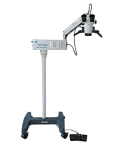
Features:
1. Japanese OLYMPUS SZ51 main microscope;
2. Excellent Achromatic Optical system;
3. Red reflex set makes surgery safer;
4. Wide field of view,solid sense and good depth of field;
5. Adopted coaxial illumination imported halogen lamp and fiber optics,with high brightness;
6. Assistance Microscope optional: 7X;
7. Imported optical system and parallel optical path design,with sharp and high resolution images;
8. Imported motor, with low noise and strong stability. -

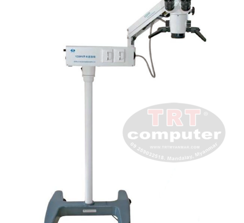
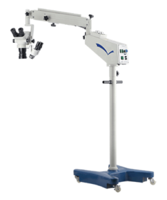
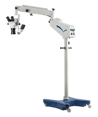
YZ20P5 Surgical Microscope is a simple binocular coaxial microscope for a single man. It is small, light weight and in high agilrly, to meet general requirements of surgery, especially suitable for mobile medical treatment.
Features:
1. Multi-layer technology is adopted in optical lens;
2. Configured with foot control focus,3 step magnifications, deep field of view, and good binocular fusion;
3. Apochrornatic technology is used to bring clearer vision Sr Me operators;
4. Light weight and compact, especially applicable for mob!, treatment;
5. Optional despoil components to make machine more portable;
6. Optional Objecfiye Lens: F250, F300, F350, F400 -
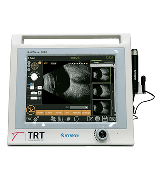
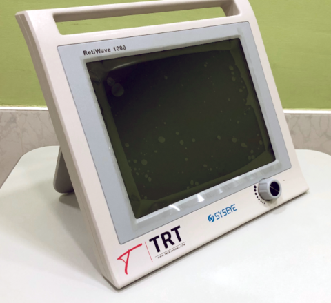
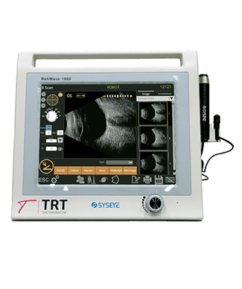
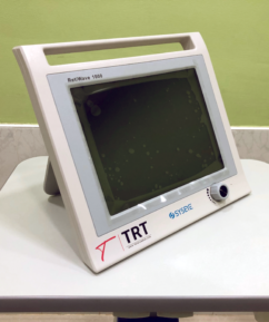
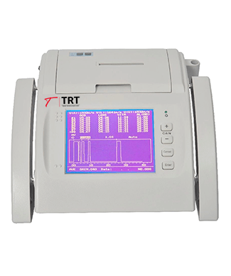
Ultrasonic A/B Scanner for Ophthalmology
1).12.1″ color and touch screen .
2). With the same software as Quantel , top one in China
With advanced intelligent digital software and parameters of freezed and Stored images could be adjusted voluntarily.
3).Can have different results on the report according to different constants A
Scan A measures data for every part of the eyeball as anterior chamber depth, lens thickness axial length and so on which are needed in ophthalmic surgeries , and calculates IOL by axial length.
4).B scanner use electromagnetic ,better save B probe life .
Scan B displays profile images of the eyeball clearly and directly. Scanning anatomical forms and nidi inside the eyeball, doctors can diagnose accurately for examination of cataract , vitreous body disease, ocular trauma, detachment of retina or choroid , macula disease , and intraocular tumor, etc
5). B scanner ,video playback of 100 images.
6). A scanner , one group with 10 data to get average ,At the same time the accuracy is 0.05mm
-
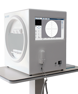
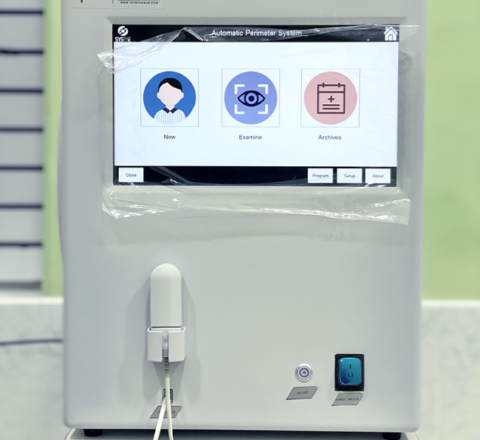
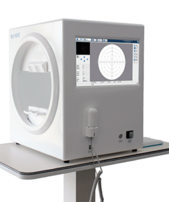
Automatic Computer Perimeter
The BIO-1000 automated perimeter absorbs the advantages of international advanced perimeter devices. It comprises the highly integrated computer, optics, machinery and electronics systems. Incorporated with the advanced configuration, comprehensive software inspection categories, and strictly in accordance with international Goldman standard, it provide scientific means for glaucoma, funds disease, visual pathway injury and neurological diseases,
– *Comprehensive real—time monitoring,
– heiji—krakau physiological blind spot monitoring,
– gaze tracking/head position tracking,
– automatic measurement of pupil diameter,
– reduce the impact of pupil effect on visual field detection.
– *Personalized design, accurate clinical analysis,
– accurate and rapid examination strategy.
– *Under international Goldman standard, providing a variety of classic test procedures and report analysis -
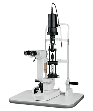
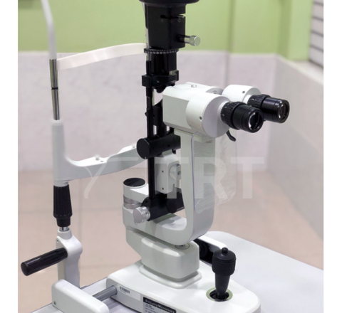
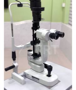
Feature
2 magnifications with 10x ,16x field of view
can get the distinct image ,also , with new illumination system
and the bigger light spot diameter ,slit width reaches 14 mm
can better observe the eye situation .
-
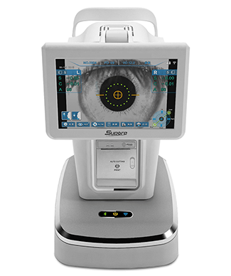
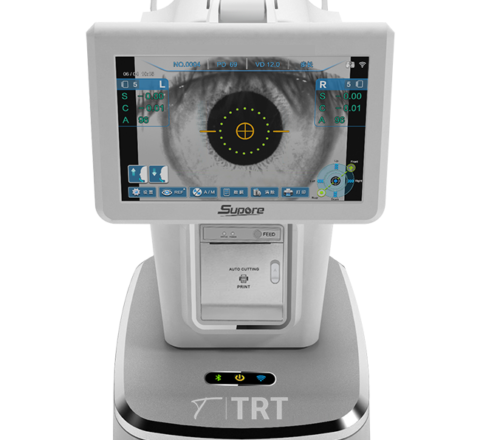
-Measuring left/right eye automatically, no need for manual switch.
-All operation can be done on the touch screen
-Touch screen can be rotated to any direction
-Operation is more fast and convenient -

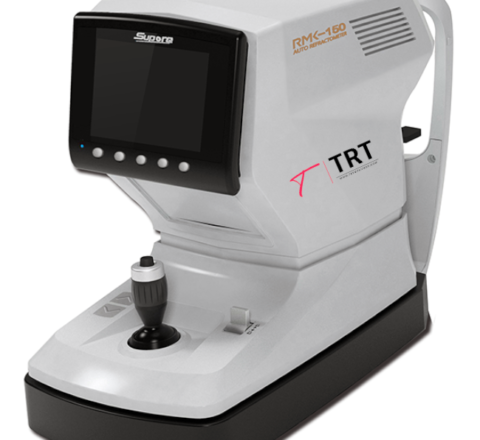
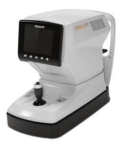
– Multiple measurement modes (SPH, CYL, CLBC, CYL&SPH)
– Automatic chin rest base and main body lifting
– Large-angle rotating screen
– Support multiple measuring functions
– Automatic fog color vision system

View Basket “RMK – 150 Auto Refractometer” has been added to your cart.